Rationale
It has long been assumed by clinicians that the shape of an aneurysm influences its risk of rupture. However, current risk prediction tools do not include detailed information from imaging data. Morphological features from vessel segmentation and haemodynamic features from computational fluid dynamic (CFD) analysis have shown differences between ruptured and unruptured aneurysms.
However, previous research is largely based on small, cross-sectional datasets which include imaging following rupture. These limitations make it difficult to apply their findings to clinical practice.
ROAR-FLOW will be the largest study of its kind, using imaging collected before rupture. By combining this with long-term follow-up, ROAR-FLOW will provide the clearest picture yet of how these features predict rupture. This will support the development of personalised decision-making for patients with unruptured aneurysms.

Aims
1. Investigate morphology – identifying parameters associated with rupture.
2. Investigate haemodynamics – identifying parameters associated with rupture.
3. Predict rupture risk – improving decisions on who should receive treatment.
Study design
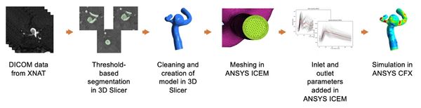
We have developed a secure, automated pipeline to transfer and anonymise imaging data from patients included in the ROAR study. Scans are sent to University Hospital Southampton via the Sectra IEP and stored in a research database (XNAT). Using a validated workflow, we will automate the analysis of aneurysm shape and blood flow using artificial intelligence (AI). Key shape and flow features will be extracted for rupture risk modelling, and explainable AI approaches will be used to group aneurysms into clusters to improve prediction.